![An Australian Government Initiative [logo]](/images/austgovt_brown_90px.gif)

Life cycle – Sporophyte development
Mosses
Where a seta is present it elongates early, while the spore capsule is still undeveloped, and the elongation is by production of additional cells. In a species with a long seta the growing sporophyte breaks through the enveloping calyptra. The lower part of the calyptra is left around the base of the seta and the calyptra's upper part is carried aloft, still covering the undeveloped spore capsule. You can see an immature sporophyte in the centre of this photo ![]() of Papillaria zeloflexicaulis. The seta has expanded and there is both a basal calyptral remnant as well as one over the apex of the sporophyte. In this species the calyptra is clearly rather hairy. By contrast, the calyptra of Encalypta vulgaris
of Papillaria zeloflexicaulis. The seta has expanded and there is both a basal calyptral remnant as well as one over the apex of the sporophyte. In this species the calyptra is clearly rather hairy. By contrast, the calyptra of Encalypta vulgaris ![]() is smooth. The upper part of the calyptra will eventually become loose and will fall off the capsule as it gets close to maturity. This photo
is smooth. The upper part of the calyptra will eventually become loose and will fall off the capsule as it gets close to maturity. This photo ![]() shows a still green but well-expanded spore capsule of Pleurophascum grandiglobum. The brown, triangular piece of tissue sitting on the capsule is the upper calyptral remnant, quite loose by now.
shows a still green but well-expanded spore capsule of Pleurophascum grandiglobum. The brown, triangular piece of tissue sitting on the capsule is the upper calyptral remnant, quite loose by now.
The seta of an immature sporophyte is not fairly straight in all species. That the seta can be twisted is shown by Funaria hygrometrica ![]() and even more so by Campylopus introflexus
and even more so by Campylopus introflexus ![]() . In the latter the immature setae are so contorted that the young spore capsules are held down amongst the leaves of the cushion composed of massed gametophyte plants. It is only near sporophyte maturity that the seta uncoils and raises the spore capsule above the moss cushion. In order to take that photograph of immature Campylopus introflexus sporophytes the moss cushion had to be teased open a little, and some plants removed, in order to reveal the spore capsules.
. In the latter the immature setae are so contorted that the young spore capsules are held down amongst the leaves of the cushion composed of massed gametophyte plants. It is only near sporophyte maturity that the seta uncoils and raises the spore capsule above the moss cushion. In order to take that photograph of immature Campylopus introflexus sporophytes the moss cushion had to be teased open a little, and some plants removed, in order to reveal the spore capsules.
The early stage of sporophyte development, where there is a seta, is often referred to as the spear stage because the undeveloped spore capsule typically shows, at most, as a slight thickening at the top of the seta and so resembles a spearhead on a spear shaft. The spore capsule will mature and enlarge atop the seta. The seta and immature capsule in the young sporophyte are both green and contain photosynthesizing cells but the sporophyte is still heavily reliant on nutrients passing to it from the gametophyte. A study into photosynthetic activity of the spore capsules of three moss species showed that the photosynthesizing capsule of Funaria hygrometrica contributes about 50% of its nutrition needs during the later stage of capsule expansion. For the species Mnium hornum the figure is about 20% and for Pleuridium acuminatum it is about 10%![]() .
.
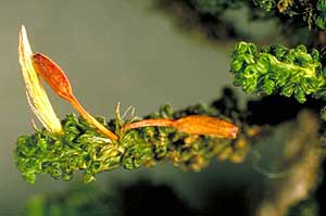 Macromitrium with sporophytes |
 Peristome teeth on moss spore capsule |
The sporophyte eventually stops photosynthesis and the capsule turns brown late in sporophyte development, as does the seta if present. Here ![]() is a colony of a species in the genus Bryum in which all the spore capsules are still immature. Amongst the setae some are green and some are already brown. It is common to see sporophytes in various stages of development. In this photo (right) of a plant of the genus Macromitrium there is one immature sporophyte, still within a yellowish, fibrous calyptra, as well as two fully mature sporophytes.
is a colony of a species in the genus Bryum in which all the spore capsules are still immature. Amongst the setae some are green and some are already brown. It is common to see sporophytes in various stages of development. In this photo (right) of a plant of the genus Macromitrium there is one immature sporophyte, still within a yellowish, fibrous calyptra, as well as two fully mature sporophytes.
In a species with no seta, or just a very short seta, it is the enlarging capsule that ruptures the calyptra. The sporophyte of Goniomitrium acuminatum ![]() has a very short seta. The photo shows some enlarged but still green spore capsules, each within the distinctive 8-pleated calyptra of this species.
has a very short seta. The photo shows some enlarged but still green spore capsules, each within the distinctive 8-pleated calyptra of this species.
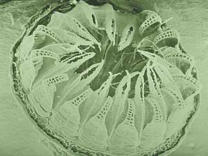
In the majority of mosses the spore capsule develops a mouth through which the spores will eventually be released. In a small number of moss genera the capsules simply disintegrate or open by means of slits, as noted in the spore DISPERSAL page. Where there is a mouth it is at the opposite side of the capsule to the point where the capsule is joined to the seta. Initially the mouth is covered by a small cap called an operculum. As the spore capsule matures and expands the upper calyptra remnant falls off. When the spores within the capsule are mature the operculum is shed. Once the operculum has been shed the mouth is exposed. In a number of moss species the mouth is surrounded by a bare rim ![]() but a greater number of species have capsules with teeth or hairs around the mouth. The teeth are called peristome teeth and, when present, there may be one ring or two rings of teeth around the margin of the mouth. This photo (right) shows an electron microscope view of a mouth with two rings of peristome teeth. Here is a side view
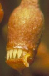
but a greater number of species have capsules with teeth or hairs around the mouth. The teeth are called peristome teeth and, when present, there may be one ring or two rings of teeth around the margin of the mouth. This photo (right) shows an electron microscope view of a mouth with two rings of peristome teeth. Here is a side view ![]() and here's a closer view at just one of the teeth
and here's a closer view at just one of the teeth ![]() . Teeth or hairs around the mouth play a role in spore DISPERSAL.
. Teeth or hairs around the mouth play a role in spore DISPERSAL.
This photo ![]() shows numerous sporophytes of Entosthodon apophysatus, viewed from above. The capsules are still green and have their calyptrae, each of which has a long beak. The calyptrae are thin and through them you can see the circular opercula. Here
shows numerous sporophytes of Entosthodon apophysatus, viewed from above. The capsules are still green and have their calyptrae, each of which has a long beak. The calyptrae are thin and through them you can see the circular opercula. Here ![]() is a side view. You can see some shed calyptrae on the ground and get a better view of the opercula of a few capsules. A couple of the capsules on the right have even shed their opercula. This final photo
is a side view. You can see some shed calyptrae on the ground and get a better view of the opercula of a few capsules. A couple of the capsules on the right have even shed their opercula. This final photo ![]() shows just a few brown sporophytes in side view.
shows just a few brown sporophytes in side view.
In many species of the family Polytrichaceae there is a circular membrane or epiphragm that is attached to the ends of short peristome teeth. This leaves just a ring of tiny gaps around the mouth through which spores can be released. The accompanying photo ![]() shows several spore capsules of Polytrichum juniperinum. You can see the white epiphragms in four and the fifth, in side view, has the operculum still attached. In this species the operculum has a central, finger like outgrowth.
shows several spore capsules of Polytrichum juniperinum. You can see the white epiphragms in four and the fifth, in side view, has the operculum still attached. In this species the operculum has a central, finger like outgrowth.
 As spore capsules mature they dry and shrink. In many species the cells of the operculum are thicker walled than those of the rest of the spore capsule and so shrink less on drying. Around the margin of the operculum is a ring of cells, called the annulus, which connects the operculum to the rest of the capsule. The cells of the annulus are large, thin-walled elastic cells. The difference in shrinking between operculum and the rest of the capsule creates tensions in the annulus which eventually breaks free and uncoils, thereby releasing the operculum. In the species Gemmabryum dichotomum shown in this photo
 ![]() the cells of the annulus in one capsule are uncoiling. Look at the three spore capsules across the centre of the photo. The one on the left still has the operculum attached, the one in the middle is losing its operculum and the one on the right has shed its operculum. Here (left) is a closer look at the uncoiling annulus of the central capsule. You can also see the peristome teeth.
the cells of the annulus in one capsule are uncoiling. Look at the three spore capsules across the centre of the photo. The one on the left still has the operculum attached, the one in the middle is losing its operculum and the one on the right has shed its operculum. Here (left) is a closer look at the uncoiling annulus of the central capsule. You can also see the peristome teeth.
The Sphagnum spore capsule is spherical while it is maturing. At maturity the body of the capsule starts to dry and contract, but the nature of the cells is such that the contraction is only horizontal and not vertical. The air inside is unable to escape and so is compressed more and more. Eventually the pressure build up inside the spore capsule becomes great enough to throw off the operculum and at the same time the spores are shot out very forcibly. A mature Sphagnum spore capsule is held up on a stalk but in this case the stalk is gametophyte tissue, not sporophyte tissue. When the spore capsule has matured a stalk grows and thereby raises the capsule. The stalk is certainly seta-like in its function but a seta develops from a fertilized egg. The gametophyte-derived stalk that raises the Sphagnum spore capsule is called a pseudopodium. In the genus Andreaea the spore capsule is also held aloft by a pseudopodium.
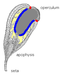 There's more than spores to a spore capsule and the internal structure can vary from species to species. The diagram (right) gives a cut-away view of a spore capsule of the moss Funaria hygrometrica, a cosmopolitan species that features commonly in structural or physiological studies.
At the bottom of the diagram you can see part of the seta. In the basal half of the capsule itself is the apophysis, also referred to as the hypophysis, and in Funaria hygrometrica ![]()
![]() the apophysis is well developed. In the centre of the apophysis is a strand of conducting tissue, the continuation of a similar strand in the seta. Surrounding this strand is spongy green tissue, with chloroplasts, and arranged somewhat palisade-like, as in the leaves of flowering plants. There are also stomata in the apophysis, thereby allowing gas exchange with the internal tissue. Spores are not produced in the apophysis but in the theca (or urn), the area between the apophysis and the mouth. The mouth is at the end opposite the seta and in this diagram it is still covered by the operculum. The proportion of the capsule taken up by the apophysis varies between species and in many species is quite rudimentary. In immature spore capsules you can often see a demarcation line or a slight change in shape, indicating the division between apophysis and theca. Many mosses have a columella, a column of sterile tissue that typically extends through the theca and which is surrounded by the spore-producing cells. The cells that will produce the spores are also referred to as the sporogenous cells or, collectively, as the archesporium. In this diagram the sporogenous cells are shown in blue, the columella is the solid area of dark grey and the cells of the annulus are in red. The yellowish areas indicate air spaces within the capsule. The size and shape of the collumella varies between species
the apophysis is well developed. In the centre of the apophysis is a strand of conducting tissue, the continuation of a similar strand in the seta. Surrounding this strand is spongy green tissue, with chloroplasts, and arranged somewhat palisade-like, as in the leaves of flowering plants. There are also stomata in the apophysis, thereby allowing gas exchange with the internal tissue. Spores are not produced in the apophysis but in the theca (or urn), the area between the apophysis and the mouth. The mouth is at the end opposite the seta and in this diagram it is still covered by the operculum. The proportion of the capsule taken up by the apophysis varies between species and in many species is quite rudimentary. In immature spore capsules you can often see a demarcation line or a slight change in shape, indicating the division between apophysis and theca. Many mosses have a columella, a column of sterile tissue that typically extends through the theca and which is surrounded by the spore-producing cells. The cells that will produce the spores are also referred to as the sporogenous cells or, collectively, as the archesporium. In this diagram the sporogenous cells are shown in blue, the columella is the solid area of dark grey and the cells of the annulus are in red. The yellowish areas indicate air spaces within the capsule. The size and shape of the collumella varies between species![]() .
.
Even when a moss seta has expanded a little and ruptured the calyptra the upper remnant on the raised immature capsule can still have an influence on capsule development. Removing the calyptra while the sporophyte is still in the spear stage leads to either cessation of capsule development or somewhat abnormal development, depending on the timing of calyptral removal. Experimental evidence has shown the effect to be physical rather than by some form of hormonal secretion![]() .
.
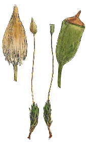
 Tayloria gunnii sporophytes |
Here is a photo of Tayloria gunnii, a species endemic to Tasmania and another moss with a well-developed apophysis. The bulk of the capsule is given over to the apophysis, the theca consisting of the conical portion above the expanded middle. Tayloria is a member of the family Splachnaceae and in that family there are many species in which the spores are dispersed by insects. In such species the stomata of mature spore capsules release insect-attracting chemicals.