![An Australian Government Initiative [logo]](/images/austgovt_brown_90px.gif)

|
|
The (macro) fungi that are dealt with in this website can be divided into two broad groups, called ascomycetes and basidiomycetes, depending on how their sexual spores are formed. All the macrofungi produce sexual spores, which result from the combination of genetic information from two parents. The function of the fruiting bodies described in the TYPES OF FUNGI SECTION is to produce and disperse sexual spores. However, many macrofungal species may also produce asexual spores - but not in such easily visible structures as those that produce the sexual spores. For the sake of brevity, the simple word "spore" will be used throughout this section but it will always mean "sexual spore", since only the sexual spores are relevant here.
In ascomycetes the spores are produced within microscopic cells called asci. The asci vary in shape from cylindric to spherical. Commonly, each ascus holds eight spores - but there are species with just one spore per ascus and others with over a hundred spores per ascus.
ASCUS DIAGRAMS HERE
In basidiomycetes the spores develop on projections that grow out from microscopic cells called basidia, rather than being enveloped within cells. In most cases the basidia are elongated and club-like, though there is variation in shape. Commonly, each basidium has four projections and four spores - but some species may have just one projection and spore per basidium and others up to eight. In most basidiomycetes the basidia have no dividing walls (or septa), but in a small number of genera the basidia are septate. The projections from the basidia are called sterigmata (singular: sterigma).
DRAWINGS OF BASIDIA: Auricularia, Tremella, Dacryopinax, 4-spored holobasidium, 6/8-spored Botryobasidium, Exidiopsis
In other websites or books you will often see the words ascospore and basidiospore used when it is necessary to emphasize the method of spore production.
Of the fruiting bodies described in the TYPES OF FUNGI section, the cup (or disk) fungi and the flask fungi are ascomycetes; the truffle-like fungi include both ascomycetes and basidiomycetes and the other fruiting bodies are basidiomycetes. While the basidiomycetes show a greater variety of fruiting body shapes, there are more ascomycete than basidiomycete species.
The features of the asci and basidia are of great importance in fungal classification and you can find out more about this in the CLASSIFICATION & IDENTIFICATION SECTION. The remainder of this section is devoted to explaining where you'll find the asci or basidia in various types of fruiting bodies.
This section will explain where you'll find the asci in a variety of ascomycete fruiting bodies. The diagrams that will help in the explanations are stylized, often with the size of the asci exaggerated (to show them more easily). Despite this, the diagrams will illustrate the nature of many of the ascomycete fruiting bodies you are likely to see.
 Jafneadelphus ferrugineus |
 Apothecium, asci in black area |
The most commonly seen larger ascomycete fruiting bodies are the ones known as the discomycetes, often called the "cup fungi". In appearance they look like shallow cups or fairly flattish disks. Jafneadelphus ferrugineus is flat, with only the margins slightly raised. Such a cup or disk shaped fruiting body is called an apothecium.
If you examined a cross-section of an apothecium under a microscope, you'd find the asci arranged vertically and making up much of the apothecium's upper surface, in the area marked in black in this simple diagram. These colours (and those in later diagrams) have no significance and are simply used to differentiate the various features. In many cases, when you slice an apothecium, you can see that the tissue near the upper surface differs in appearance from the tissue elsewhere in the apothecium. Sometimes this is easy to see with the naked eye, but at other times you'll need a hand lens. At a magnification of about a hundred times you can see that the surface layer clearly consists of vertically aligned elements, but you may not be able to make out the detail.
The next diagram shows the view of a part of that black area at a magnification
of about a couple of hundred times. The spores are shown as greyish blue spots
within the asci (coloured dull yellow).  Most
ascomycete species have eight spores per ascus. Between the asci are the paraphyses
(shown here as brown-walled with light grey interiors). Sometimes these are
just simple, unmodified hyphae - but often they are modified hyphae. They may
have broader apices, perhaps coloured or encrusted. This diagram represents
a vertical cross-section of a small region in the upper part of an apothecium.
The hyphae from which the asci arise are shown in darker grey.
Most
ascomycete species have eight spores per ascus. Between the asci are the paraphyses
(shown here as brown-walled with light grey interiors). Sometimes these are
just simple, unmodified hyphae - but often they are modified hyphae. They may
have broader apices, perhaps coloured or encrusted. This diagram represents
a vertical cross-section of a small region in the upper part of an apothecium.
The hyphae from which the asci arise are shown in darker grey.
As noted above, the structure of the tissue in the brown areas differs markedly from that of the layer containing the asci and paraphyses. There's more about the brown area in the CLASSIFICATION/IDENTIFICATION SECTION
Sometimes the apothecia can become slightly distorted when they get large,
as is shown by this Peziza apothecium growing on a carpet ![]() .
It has curved back noticeably.
.
It has curved back noticeably.
 |
 Leotia lubrica |
The fruiting bodies of Leotia lubrica are a few centimetres tall and,
at first glance, could be mistaken for small-capped mushrooms. However, there
are no gills under the cap and Leotia lubrica is in fact an ascomycete.
The asci (and paraphyses) are on the top of the cap, in the area marked in black
in this diagram of a cross-section. The interior of the fruiting body is hollow.
In effect, the fruiting body consists of an apothecium that has been given a
slight outward curve and then stuck on top of a short stalk.
 |
Trichoglossum walteri |
The fruiting bodies of species in the genera Geoglossum ![]() and Trichoglossum are blackish brown to black and are mostly club-like
- consisting of an expanded upper portion atop a narrower stem. They are rubbery
in texture, found on the soil and are easily overlooked since they are usually
under a centimetre at their greatest width and no more than a few centimetres
tall. The photo shows an example of Trichoglossum walteri. At first you
might think this to be one of the simple-stalked coral fungi - but Geoglossum
and Trichoglossum are ascomycetes. A cross-section of one of these fruiting
bodies would resemble this diagram. Once again, the black band represents the
area containing the asci and paraphyses. In essence, the structure is still
that of an apothecium - though much further removed from the basic disk shape
than even Leotia is. You could think of the fruiting body as an apothecium
that has been put on top of a pole, and then had the edges pulled down until
the greater part of the pole is covered.
and Trichoglossum are blackish brown to black and are mostly club-like
- consisting of an expanded upper portion atop a narrower stem. They are rubbery
in texture, found on the soil and are easily overlooked since they are usually
under a centimetre at their greatest width and no more than a few centimetres
tall. The photo shows an example of Trichoglossum walteri. At first you
might think this to be one of the simple-stalked coral fungi - but Geoglossum
and Trichoglossum are ascomycetes. A cross-section of one of these fruiting
bodies would resemble this diagram. Once again, the black band represents the
area containing the asci and paraphyses. In essence, the structure is still
that of an apothecium - though much further removed from the basic disk shape
than even Leotia is. You could think of the fruiting body as an apothecium
that has been put on top of a pole, and then had the edges pulled down until
the greater part of the pole is covered.
Each of the fruiting bodies described above could be thought of as a single apothecium, albeit highly distorted in some cases. There are some ascomycetes which look as though they have been built up from a number of apothecia.
Cytaria and Morchella
 Cyttaria |
 Cyttaria gunnii |
Both Morchella elata <<Murray;s slide>> and Cyttaria
gunnii have more complicated fruiting bodies, composed of numerous depressions
and ridges. In effect, each depression is like an individual apothecium so that,
superficially, it looks as if a number of separate apothecia (sometimes distorted
in shape) have been glued together onto a stalk (Morchella) or into a
ball (Cyttaria). The following diagrams show vertical cross-sections
of Morchella and Cyttaria. The interiors are empty, the areas
bearing asci and paraphyses are shown in black and the rest of the tissue of
the fruiting bodies is shown in brown.
 |
 Nectria fruiting bodies |
 Perithecia on dead wood |
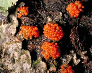
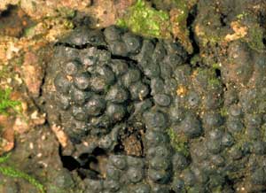
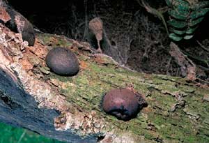
These ascomycetes differ markedly from the types so far described. The Flask
Fungi produce their spores in tiny, generally spherical, chambers (called perithecia)
which are usually under a couple of millimetres in diameter. The first photo
shows a number of tiny orange balls, each only about a millimetre or two in
diameter. Each such ball is a separate perithecium and is the fruiting body
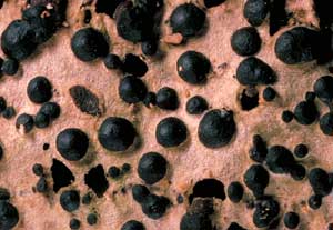
of a species of Nectria. Here's another photo showing some black hemispheres
(about 2mm in diameter) on a piece of dead wood. Once again these are examples
of perithecia. If you cross-sectioned a perithecium you'd see the asci inside,
as shown by this diagram.
Of course, this is a very simplistic diagram and there is considerable variation in both the shapes and contents of perithecia. These diagrams show some of the variation in shapes that can be seen in different species.
In some species the chamber may be empty, others may have paraphyses
or even have the entire chamber filled. The asci may be found only at the base
of the perithecium - or up the sides as well. Two examples are shown here (with
the asci in red and paraphyses in grey)
 Hypoxylon
sp. an example of communal perithecia. Hypoxylon
sp. an example of communal perithecia.
|
|
Just as there are "compound apothecial" fruiting bodies (such as Morchella and Cyttaria), so there are also "compound perithecial" fruiting bodies, where a number of perithecia are combined within a larger structure. In many perithecial ascomycetes the perithecia are embedded within a communal tissue (called a stroma, with the plural stromata). In many cases the stroma forms a dark-coloured, brittle sheet on wood as shown in this photo of Hypoxylon sp. The little pimples that dot the black sheet are the very tips of the perithecia. In cross-section such a compound fruiting body would have the following structure.The mass of dark grey represents the stroma in which the perithecia are embedded and you can see that the apical openings just protrude above the general level of the stroma.
 |
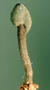
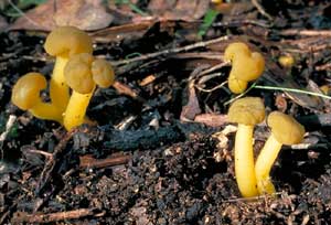
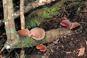
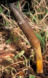 Cordyceps gunnii |
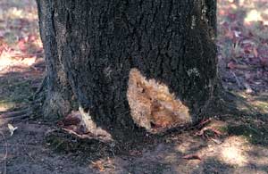
As well as such sheet-like species there are various species with markedly
three-dimensional stromata. The species of Cordyceps are parasites, mostly
of invertebrates and commonly of invertebrate larvae. The fruiting body in this
photo (of Cordyceps gunnii, ) has emerged from such a parasitized larva
in the ground ![]() .
In cross-section the fruiting body would resemble the following diagram, with
the perithecia just under the surface of the stroma (again shown in grey). The
sterile stem is shown in khaki-green. Several species of Xylaria
.
In cross-section the fruiting body would resemble the following diagram, with
the perithecia just under the surface of the stroma (again shown in grey). The
sterile stem is shown in khaki-green. Several species of Xylaria ![]() have a similar club-like appearance and internal structure (though without the
well-differentiated stem of Cordyceps). However, while these club-like
fruiting bodies are of similar shape to those of genera such as the previously
mentioned Geoglossum and Trichoglossum (and Xylaria is
even black), the internal constructions are quite different. Remember that Geoglossum
and Trichoglossum have no perithecia but have a palisade of asci and
paraphyses lining their surfaces.
have a similar club-like appearance and internal structure (though without the
well-differentiated stem of Cordyceps). However, while these club-like
fruiting bodies are of similar shape to those of genera such as the previously
mentioned Geoglossum and Trichoglossum (and Xylaria is
even black), the internal constructions are quite different. Remember that Geoglossum
and Trichoglossum have no perithecia but have a palisade of asci and
paraphyses lining their surfaces.
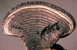
Another example of a perithecial ascomycete with a three-dimensional stroma is Daldinia concentrica, which produces hemispherical fruiting bodies (up to several centimetres in diameter) on dead wood. One photo shows a cross-section, which is also illustrated in this diagram - with the perithecia around the periphery.
 |
 Daldinia concentrica (left) diagram (above) section through fruiting body (right) |
 |
 |
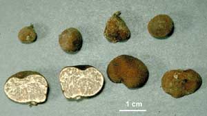
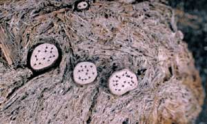 Poronia erici growing on dung |
You may often see small, black-spotted, whitish disks on herbivore dung, such
as shown in this photo. These are the fruiting bodies of a perithecial ascomycete
called Poronia erici and may grow to about a centimetre across. The black
dots are the tips of the embedded perithecia and a cross-section would show
a view much as the one in this diagram, with the perithecia under the upper
surface of the grey stroma. The khaki green represents the surrounding dung.
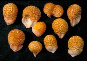
In the ascomycete truffle-like fungi the asci may be spread throughout the interior of the fruiting body, embedded in firm tissue or be lined up along the walls of internal chambers. This unidentified species of Tuber , collected in the Australian National Botanic Gardens, has one large spore per ascus, with the asci embedded in the firm internal tissue. The photo shows thin, white veins around areas that are slightly darker and glassy. Those latter areas hold the asci, which are mostly spherical but with a short, tapering "tail". The spherical spores have a net-like surface ornamentation over their surfaces and, diagrammatically, the interior looks a bit like this.
 |
|
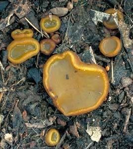
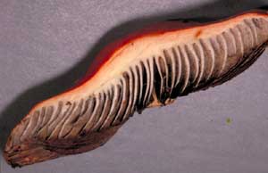
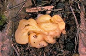 Peziza whitei |
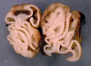 Section through Peziza whitei |
On the other hand, Peziza whitei shows a much more open appearance when
cut in half and the asci line the various convoluted surfaces. This almost brings
us back to where we started, for in some ways, you could look at this fruiting
body as simply an apothecium that has been greatly ![]() twisted
and folded in various directions.
twisted
and folded in various directions.
Basidia: where to find them
This section will explain where you'll find the basidia in a variety of the basidiomycete fruiting bodies. The diagrams that will help in the explanations are stylized, often with the size of the basidia exaggerated (to show them more easily). However, the diagrams will illustrate the nature of many of the basidiomycete fruiting bodies you are likely to see.
Mushrooms
Mushrooms are the best known basidiomycetes, so let's start with a mushroom and look at it's structure, at three different levels of magnification. Suppose you slice vertically across the outer part of a mushroom cap and pull away the small off-cut. The following diagram shows what you'd see.
 |
 Section through Hypholoma arantiacum cap |
For interest's sake, the diagram represents a mushroom with concentric rings of short, black scales on the cap. In most mushrooms the gills are V-shaped in cross-section, with the point of the V at the bottom edge of the gill. The grey colour represents the exposed flesh of the cap and gills. In some cases you'd need to use a hand lens, that magnifies 10 to 20 times, to clearly see the V shape of the gill cross-section.
The gills of a mushroom are lined with basidia. So, if you took a very thin slice, across several gills, from that exposed section, and viewed it at about a hundred times magnification you'd see the basidia sticking out from the gills, as shown on the left - though in most mushrooms the top of the V would be much narrower than shown here. The basidia are drawn in green, the spores in brown and the general gill tissue again in grey. These colours (and those in later diagrams) have no significance and are simply used to differentiate the various structures. The basidia protrude from one gill into the airspace between that gill and the neighbouring gill. During their development, the spores are exposed to the air between the gills - quite unlike the ascomycetes, where the spores are enclosed in asci.
If you took a small section from a gill (such as the area contained in the blue rectangle in the above figure) and looked at it under the microscope at a magnification of several hundred times, you'd see something resembling the figure on the right. You can now see the basidia much more clearly and also see the hyphae that make up the gill tissue. In this diagram the hyphae are marked by the parallel pairs of greyish-blue lines and you can also see the septa that divide the filamentous hyphae into separate compartments. Notice that the basidia are the terminal elements of various hyphae and that between the basidia there are numerous ordinary hyphae. You can follow the hyphae back from the basidia and see that they bend upwards and may weave under and over each other.
There is much variation in shape of basidia and arrangement of hyphae in the gills. Moreover, in some species, there may be other structures (such as cystidia SEE CLASSIFICATION & IDENTIFICATION SECTION) also protruding from the gill surface. Similar comments hold for the other types of fruiting bodies discussed below.
As noted above, the V-shape of the gill cross section was exaggerated in the diagrams. The photo shows a slice taken from the cap of a mushroom of Hypholoma aurantiaca . You can see the white flesh that makes up the relatively narrow V-shape in each of the gill cross-sections. The gill faces have numerous basidia bearing dark brown spores - hence the dark coloured areas on either side of each gill section.
Boletes
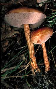 Austroboletus sp. |
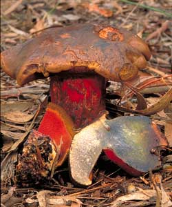 A bolete showing sectional views |
In some ways the boletes are very mushroom-like. A bolete is a fleshy fruiting body consisting of a cap on a stem - but with a spongy pore layer (rather than gills) on the underside of the cap. In this photo of an undescribed species of Austroboletus, you can see some of that spongy, pored layer. You can imagine each pore to be the mouth of a short, vertical tube. Thus, the area beneath the cap superficially resembles a mass of closely-packed tubes, and an enlarged view of a small section of the underside of the cap is shown schematically in this diagram.
The inner walls of those tubes are lined with basidia. The walls themselves are composed of hyphae, in the same way as the body of a mushroom gill. The photo (above right) shows a bolete that has been cut in half vertically. The half on the left shows a view of the underside with its tiny, red pores. The half on the right shows that the white flesh of the cap and stem has blued on exposure to the air. You can also see a side-on view of the vertically sliced tubes that constitute the spore-producing area below the flesh of the cap.
Polypores
The polypores are a little like boletes, in that they also have pores that
are mouths of short, vertical tubes. Once again the inner walls of the tubes
are lined with basidia. The fruiting bodies have varied shapes. Some, like Polyporus
arcularius, consist of a cap on a stem but most polypores are either bracket-like
in form (such as Ganoderma australe ![]() and Phaeotrametes decipiens or, like Macrohyporia dictyopora ,
more-or-less sheet-like and pressed closely to the underlying wood.
and Phaeotrametes decipiens or, like Macrohyporia dictyopora ,
more-or-less sheet-like and pressed closely to the underlying wood.
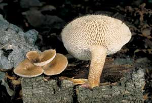 Polyporus arcularius |
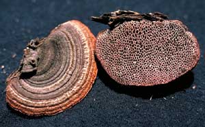 Phaeotrametes decipiens |
 Macrohyporia dictyopora |
Corticioid, stereoid and coral fungi
 basidia lining the undersides of the fruiting bodies |
The corticioid and stereoid fungi have basidia lining the undersides of the fruiting bodies. The corticioid fungi have sheet-like fruiting bodies - often smooth but also found with minor bumps, ridges, spines, etc on the otherwise two-dimensional sheets. The stereoid fruiting bodies also have generally smooth undersides but are bracket-like or with a stem - and therefore differ from the corticioid fungi by being markedly three-dimensional. The following diagram, represents a corticoid fruiting body (the brighter brown) growing on the underside of a piece of wood lying on the ground. You can see the basidia protruding into the air.
|
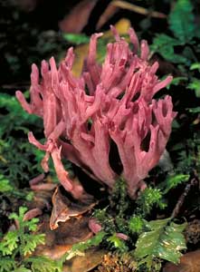
Clavaria zollingeri (right) |
 |
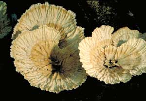
A funnel-shaped stereoid fungus such as Cymatoderma elegans var. lamellatum has the basidia on the underside of the sloping funnel area.
In a coral fungus, such as Clavaria zollingeri the basidia are found on many of the branches, as shown in this diagram.
A couple of gelatinous fungi with septate basidia
|
Tremella (right) |
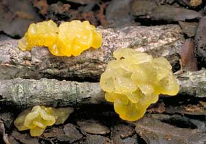 |
The irregularly shaped jelly fungi (such as the species of Tremella) have the basidia in the convoluted surfaces of the fruiting bodies. As shown in the CLASSIFICATION SECTION, the basidia of the jelly fungi are septate along their long axes and have long, often weakly sinuous sterigmata. In contrast to the fungi described above, the basidia of the jelly fungi are within the gelatinous fruiting body, with only the ends of the sterigmata (and the spores) protruding into the air. The basidia and hyphae that make up the fruiting bodies are often embedded within a gelatinous matrix that can make the hyphae hard to see. In this diagram the dull yellowish colour represents the gelatinous matrix and the thick, grey, wavy lines are the hyphae of the fruiting body. As before, the basidia and spores are coloured green and brown respectively.
 basidia in smooth, semi-glossy undersurface |
 Auricularia sp. |
The fresh fruiting bodies of species in the genus Auricularia are gelatinous and somewhat ear-shaped (hence the common name of Wood Ears, for they grow on wood). The basidia are elongated, tapering at their ends and septate across their width. As in the jelly fungi, the basidia of Auricularia are within the fruiting bodies with just the ends of the sterigmata (and the spores) beyond the surface. The basidia are in the smooth, semi-glossy undersurface - not the dull, roughened to bristly upper surface. This diagram shows the arrangement in Auricularia, using the same colour code as in the preceding jelly fungus diagram.
Truffle-like fungi
 Setchelliogaster sp. (right) |
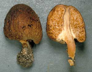 |
The fruiting bodies of the basidiomycete truffle-like fungi are varied in form, sometimes stalked but mostly stalk-less and more-or-less spherical in shape. Internally the fruiting bodies are chambered, with the chambers of some species easy to see with the naked eye - but a hand lens is needed to see the individual chambers in other species. The basidia line the walls of the chambers and protrude into the interiors of the chambers - which are empty in many species, but not in all. In shape the chambers may be anything from spherical to quite contorted. This diagram represents the cross-section of a truffle-like fruiting body. The solid, fleshy areas of the truffle are coloured brown and you can see the basidia (coloured green and bearing dark brown spores) lining the walls of those chambers. Of course, this diagram exaggerates the sizes of both the basidia and the chambers and this simplistic two-dimensional figure does not do justice to the internal convolutions of three-dimensional fruiting body. The photograph of the stalked Setchelliogaster fruiting body may give you a better idea of the internal structure. On the left you can see the outside appearance and on the right the internal structure. In the latter you can see some of the chambers (of varied forms) and get an idea of the intricate way in which the internal tissue creates those chambers.
Gasteromycetes
In all of the preceding basidiomycetes the basidia are persistent. Even by the time a mushroom has largely rotted away you can usually still find basidia on the gills. There are several common groups of basidiomycetes where the mature fruiting body has spores - but no basidia. To find the basidia you must have an immature (but not too young!) fruiting body, because once the spores have been produced the basidia break down. Examples of such fungi are various powdery spored fruiting bodies (the puffballs and similar fungi), the slimy-spored stinkhorns and the Bird's Nest Fungi.
These are examples of a subgroup of the basidiomycetes commonly called the gasteromycetes. The word gasteromycete literally means "stomach fungus" - and these fungi produce their spores inside the fruiting body that, at least initially, is enclosed within an outer skin. While, as a group, these fungi may all be referred to as gasteromycetes, that name does not signify close evolutionary relationships between all the members. The name is simply one of convenience - rather than being one of classificatory importance. Reflecting their diverse origins, these fungi show considerable variation in both the overall appearance and internal structure of the immature, basidia-bearing fruiting bodies. Many start as more-or-less spherical masses of homogenous tissue, and then develop various patterns of internal spore-bearing areas. In some species these spore-bearing areas consist of empty cavities, with the basidia lining the walls and protruding into the empty interiors. In others the spore bearing areas may be filled with basidia and hyphae or a gelatinous matrix while the spores are developing.
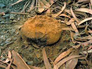
Pisolithus is a common powdery-spored gasteromycete genus, with the fruiting bodies often seen pushing up through bitumen roads.
 |
cross-section (right) |
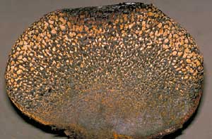 |
A vertical cross-section of such a fruiting body shows a powdery region at the top, but with the lower part looking as though it consisted of a multitude of densely packed rice grains (called peridioles), comprising packets of spores. The peridioles are relatively thin skinned and the whole mass is contained in a brittle outer skin. This highly stylized diagram shows a vertical cross-section through a very immature Pisolithus fruiting body. The black circle represents the outer skin, the internal tissue is in brown and there are numerous chambers (shown in white). Each of these chambers eventually becomes the interior of a peridiole. These chambers are isolated from each other and contain the basidia. The chambers fill quickly with a gelatinous matrix, the spores become detached from their basidia and complete their development (after detachment) within the gelatinous interior. Of course, in most cases people only see the Pisolithus fruiting bodies in the advanced, powdery stage as shown in the photograph.
 Cyathus novae-zealandiae (right) |
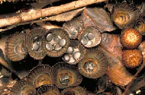 |
The mature fruiting body of a Bird's Nest Fungus (such as this Cyathus novae-zealandiae) consists of a leathery, cup-like structure that contains a number of disc-shaped "eggs" (peridioles again). These peridioles differ from the Pisolithus peridioles by being enclosed in a thick outer casing and by becoming separated from each other. The cup-like structure is usually no more than a centimetre in diameter at the top. Before the Cyathus fruiting bodies are mature, they have a membrane across the top of the cup. You can see a couple of immature fruiting bodies at the right in the photograph. The following diagram shows an immature cup, cut away to show it full of peridioles. The figure on the right shows the basidia in the central part of one such peridiole. The thick brown line represents the peridiole's hard outer casing. At a young stage, the interior of a peridiole has a gelatinous filling and at an even younger stage the fruiting body would have had the same type of chambered appearance as shown in the stylized diagram for Pisolithus.
The preceding cross-section diagram shows a very simple type of internal structure, but numerous gasteromycetes have much more complicated patterns of development.
 Many
mature puffballs have a very simple structure. There's just a thin-walled sack
of powdery spores with a hole at the top, through which the spores are puffed
out when the sack is compressed. This simplicity at maturity belies the complex
internal structure of the immature fruiting body during spore production. At
that stage the fruiting body has a very convoluted interior. The diagram represents
a stylized vertical cross-section of such a young puffball. Here the cavities
are not formed as separate chambers, but within ingrowths of tissue from the
periphery. The basidia develop on those convoluted surfaces, shown here in brown.
Many
mature puffballs have a very simple structure. There's just a thin-walled sack
of powdery spores with a hole at the top, through which the spores are puffed
out when the sack is compressed. This simplicity at maturity belies the complex
internal structure of the immature fruiting body during spore production. At
that stage the fruiting body has a very convoluted interior. The diagram represents
a stylized vertical cross-section of such a young puffball. Here the cavities
are not formed as separate chambers, but within ingrowths of tissue from the
periphery. The basidia develop on those convoluted surfaces, shown here in brown.
 See
colour coding See
colour codingin diagram to left of page. |
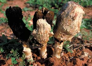
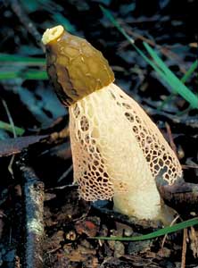 Dictyophora multicolor |
The fruiting bodies of stinkhorns in the genera Dictyophora, Mutinus
and Phallus consist of a stem with the slimy spore mass on a small cap
at the top of the stem. The cap isn't spread out (like a typical mushroom) but
hugs the stem fairly closely. In this picture of Dictyophora multicolor
you can see the small, slimy cap. In addition, there is a mesh-like skirt that
hangs down below the cap. This picture of a dissected Phallus rubicundus
![]() shows the cap a little more clearly.
shows the cap a little more clearly.
 In
line with the style of the two gasteromycete diagrams above, the following diagram
represents a vertical cross-section of a young stinkhorn from any of the three
genera listed above. You can see the unexpanded stem, the curved cap and the
short projections (growing in from the marginal tissue) that bear the basidia.
In
line with the style of the two gasteromycete diagrams above, the following diagram
represents a vertical cross-section of a young stinkhorn from any of the three
genera listed above. You can see the unexpanded stem, the curved cap and the
short projections (growing in from the marginal tissue) that bear the basidia.
 It
is important to realize that, as in the case of the truffle-like diagram above,
these gasteromycete diagrams are also very simplistic, two-dimensional representations
of considerable three-dimensional complexity. Looking more closely at the young
stage of Dictyophora, here is a more detailed (but still stylized) diagram
of the structure, showing an enlarged view of the area contained within the
blue rectangle above. The young stinkhorn is enclosed within a thin, leathery
skin, represented here by the thin dark-grey line. Immediately beneath that
skin is a relatively broad gelatinous zone (the stippled band). Next are the
finger-like to plate-like projections (khaki green) that bear the basidia; the
body of the eventual cap (bluish); the tissue that will form the skirt (shown
here as a red net); other hyphal/gelatinous tissue (stippled pale grey) and
the stem (the brown vertical bar at the right). The projections can form quite
intricate channels and chambers but (once the spores have matured) they break
down into the smelly, khaki to brown slime that holds the spores on the outside
of the cap of the mature fruiting body.
It
is important to realize that, as in the case of the truffle-like diagram above,
these gasteromycete diagrams are also very simplistic, two-dimensional representations
of considerable three-dimensional complexity. Looking more closely at the young
stage of Dictyophora, here is a more detailed (but still stylized) diagram
of the structure, showing an enlarged view of the area contained within the
blue rectangle above. The young stinkhorn is enclosed within a thin, leathery
skin, represented here by the thin dark-grey line. Immediately beneath that
skin is a relatively broad gelatinous zone (the stippled band). Next are the
finger-like to plate-like projections (khaki green) that bear the basidia; the
body of the eventual cap (bluish); the tissue that will form the skirt (shown
here as a red net); other hyphal/gelatinous tissue (stippled pale grey) and
the stem (the brown vertical bar at the right). The projections can form quite
intricate channels and chambers but (once the spores have matured) they break
down into the smelly, khaki to brown slime that holds the spores on the outside
of the cap of the mature fruiting body.
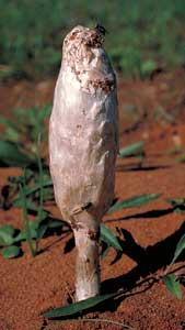 |
 Podaxis pistillaris |
The stem-and-cap type of development (as shown above in the simple brown diagram) is not restricted to stinkhorns. The powdery spored Podaxis pistillaris also has such a form when young. In the mature stage Podaxis fruiting bodies look like powdery drumsticks. The powdery region is initially contained within a brittle, white outer skin, but the skin breaks away to expose the dark brown to black spore powder.
There are a few more forms of immature gasteromycete fruiting bodies than shown here, but the types shown here will give you some idea of the variety. As you can see, the gasteromycete fruiting bodies can be quite complex and to understand the complexity you need to follow the development of the fruiting body and, in particular, note where the basidia form.