![An Australian Government Initiative [logo]](/images/austgovt_brown_90px.gif)

Two Groups - classifying fungi into ascomycetes and basidiomycetes:
The cup fungi - and relatives
 Jafneadelphus ferrugineus |
 Apothecium, asci in black area |
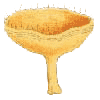
The most commonly seen larger ascomycete fruiting bodies are the ones known as the discomycetes, often called the "cup fungi". In appearance they look like shallow cups or fairly flattish disks. Jafneadelphus ferrugineus is flat, with only the margins slightly raised. Such a cup or disk shaped fruiting body is called an apothecium.
If you examined a cross-section of an apothecium under a microscope, you'd find the asci arranged vertically and making up much of the apothecium's upper surface, in the area marked in black in this simple diagram. These colours (and those in later diagrams) have no significance and are simply used to differentiate the various features. In many cases, when you slice an apothecium, you can see that the tissue near the upper surface differs in appearance from the tissue elsewhere in the apothecium. Sometimes this is easy to see with the naked eye, but at other times you'll need a hand lens. At a magnification of about a hundred times you can see that the surface layer clearly consists of vertically aligned elements, but you may not be able to make out the detail.
The next diagram shows the view of a part of that black area at a magnification
of about a couple of hundred times. The spores are shown as greyish blue spots
within the asci (coloured dull yellow). 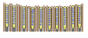 Most
ascomycete species have eight spores per ascus. Between the asci are the paraphyses
(shown here as brown-walled with light grey interiors). Sometimes these are
just simple, unmodified hyphae - but often they are modified hyphae. They may
have broader apices, perhaps coloured or encrusted. This diagram represents
a vertical cross-section of a small region in the upper part of an apothecium.
The hyphae from which the asci arise are shown in darker grey.
Most
ascomycete species have eight spores per ascus. Between the asci are the paraphyses
(shown here as brown-walled with light grey interiors). Sometimes these are
just simple, unmodified hyphae - but often they are modified hyphae. They may
have broader apices, perhaps coloured or encrusted. This diagram represents
a vertical cross-section of a small region in the upper part of an apothecium.
The hyphae from which the asci arise are shown in darker grey.
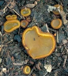
Sometimes the apothecia can become slightly distorted when they get large,
as is shown by this Peziza apothecium growing on a carpet ![]() .
It has curved back noticeably.
.
It has curved back noticeably.
 |
 Leotia lubrica |
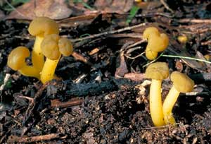
The fruiting bodies of Leotia lubrica are a few centimetres tall and,
at first glance, could be mistaken for small-capped mushrooms. However, there
are no gills under the cap and Leotia lubrica is in fact an ascomycete.
The asci (and paraphyses) are on the top of the cap, in the area marked in black
in this diagram of a cross-section. The interior of the fruiting body is hollow.
In effect, the fruiting body consists of an apothecium that has been given a
slight outward curve and then stuck on top of a short stalk.
 |
Trichoglossum walteri |
The fruiting bodies of species in the genera Geoglossum ![]() and Trichoglossum are blackish brown to black and are mostly club-like
- consisting of an expanded upper portion atop a narrower stem. They are rubbery
in texture, found on the soil and are easily overlooked since they are usually
under a centimetre at their greatest width and no more than a few centimetres
tall. The photo shows an example of Trichoglossum walteri. At first you
might think this to be one of the simple-stalked coral fungi - but Geoglossum
and Trichoglossum are ascomycetes. A cross-section of one of these fruiting
bodies would resemble this diagram. Once again, the black band represents the
area containing the asci and paraphyses. In essence, the structure is still
that of an apothecium - though much further removed from the basic disk shape
than even Leotia is. You could think of the fruiting body as an apothecium
that has been put on top of a pole, and then had the edges pulled down until
the greater part of the pole is covered.
and Trichoglossum are blackish brown to black and are mostly club-like
- consisting of an expanded upper portion atop a narrower stem. They are rubbery
in texture, found on the soil and are easily overlooked since they are usually
under a centimetre at their greatest width and no more than a few centimetres
tall. The photo shows an example of Trichoglossum walteri. At first you
might think this to be one of the simple-stalked coral fungi - but Geoglossum
and Trichoglossum are ascomycetes. A cross-section of one of these fruiting
bodies would resemble this diagram. Once again, the black band represents the
area containing the asci and paraphyses. In essence, the structure is still
that of an apothecium - though much further removed from the basic disk shape
than even Leotia is. You could think of the fruiting body as an apothecium
that has been put on top of a pole, and then had the edges pulled down until
the greater part of the pole is covered.
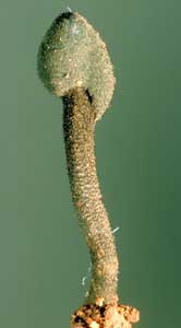
Each of the fruiting bodies described above could be thought of as a single apothecium, albeit highly distorted in some cases. There are some ascomycetes which look as though they have been built up from a number of apothecia.
Cytaria and Morchella
 Cyttaria |
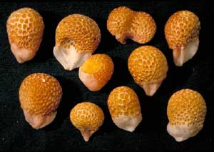 Cyttaria gunnii |
Both Morchella elata and Cyttaria gunnii have more complicated
fruiting bodies, composed of numerous depressions and ridges. In effect, each
depression is like an individual apothecium so that, superficially, it looks
as if a number of separate apothecia (sometimes distorted in shape) have been
glued together onto a stalk (Morchella) or into a ball (Cyttaria).
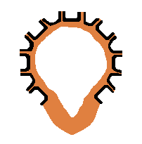
The diagrams show vertical cross-sections of Morchella and Cyttaria.
The interiors are empty, the areas bearing asci and paraphyses are shown in
black and the rest of the tissue of the fruiting bodies is shown in brown.
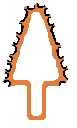 |
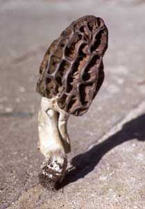 Morchella elata |